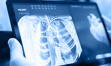
Improved Pulmonary Embolism Detection in CT Pulmonary Angiogram Using Hybrid Vision Transformers and Deep Learning Techniques
Pulmonary embolism (PE) is one of the leading causes of cardiovascular mortality worldwide, ranking only behind myocardial infarction and stroke. Despite major advances in medical imaging, timely diagnosis of PE remains a significant clinical challenge. CT Pulmonary Angiography (CTPA) is considered the gold standard for diagnosing pulmonary embolism; however, interpreting a single CTPA examination often requires radiologists to review hundreds of image slices, making the process time-consuming, cognitively demanding, and susceptible to diagnostic variability.
Recent advances in Artificial Intelligence (AI), Deep Learning, and Computer Vision have opened new opportunities to improve the speed and accuracy of pulmonary embolism detection. Vision Transformers, Convolutional Neural Networks (CNNs), and ensemble learning techniques are transforming medical image analysis by enabling automated recognition of subtle radiological patterns that may be overlooked during routine clinical interpretation.
In this research presentation, Abeer Abdelhamid, PhD Researcher at the Department of Electronics and Communications Engineering, Faculty of Engineering, Mansoura University, and Assistant Lecturer at the Higher Technological Institute of Applied Health Science, presents a comprehensive portfolio of her published research focusing on AI-powered pulmonary embolism detection.
Rather than presenting a single study, this video showcases a series of complementary research projects that investigate multiple artificial intelligence architectures for pulmonary embolism detection from CT Pulmonary Angiography scans and structured clinical data. The presented work combines state-of-the-art Vision Transformers, Deep Learning ensembles, attention mechanisms, diffusion pretraining, and explainable artificial intelligence to create highly accurate computer-aided diagnostic systems capable of supporting radiologists in clinical decision-making.
Collectively, these studies demonstrate how modern AI techniques can significantly improve diagnostic performance while providing greater interpretability, robustness, and scalability for real-world clinical applications.
Research Overview
This research presentation summarizes several internationally published studies that investigate different computational strategies for pulmonary embolism detection using advanced machine learning and deep learning methodologies.
The research explores multiple complementary diagnostic perspectives, including:
-
- Image-based pulmonary embolism detection using Hybrid Vision Transformers.
- Ensemble Deep Learning models combining CNNs and Transformer architecture.
- Diffusion-pretrained CNN-Transformer networks operating on complete 3D CTPA volumes.
- Clinical metadata analysis using optimized stacking ensemble learning.
- Explainable AI techniques that enhance transparency and clinical trust.
- Attention-based architectures for accurate pulmonary embolism localization and classification.
Unlike conventional AI systems that rely on a single predictive model, this body of work investigates multiple independent yet complementary frameworks capable of analyzing pulmonary embolism from slice-level, image-level, volume-level, and patient-level perspectives.
The presented research addresses one of the major limitations of current computer-aided diagnosis systems: balancing high diagnostic accuracy with model interpretability while maintaining clinical applicability.
What You Will Learn in This Video
This research provides an in-depth overview of how artificial intelligence is transforming pulmonary embolism diagnosis through advanced medical image analysis.
Throughout the presentation, viewers will gain insights into:
-
- The clinical challenges associated with pulmonary embolism diagnosis.
- Why CTPA interpretation remains difficult despite modern imaging technology.
- The limitations of traditional convolutional neural networks in PE detection.
- How Vision Transformers improve global contextual understanding of medical images.
- The integration of Discrete Wavelet Transform (DWT), Sobel edge enhancement, Autoencoders, and Deep Learning for feature extraction.
- The advantages of ensemble learning over single-model classification.
- Diffusion-based self-supervised pretraining for medical imaging.
- Attention mechanisms that identify clinically relevant embolic regions.
- Explainable AI approaches include SHAP and attention visualization.
- Future directions for multimodal AI-assisted pulmonary embolism diagnosis.
The presentation also illustrates how combining multiple complementary AI models can produce more reliable diagnostic systems capable of supporting radiologists in routine clinical practice.
Key Research Contributions
The research portfolio introduces several innovative contributions to the field of artificial intelligence in medical imaging.
Hybrid Vision Transformer Framework
A novel diagnostic pipeline combining Autoencoders, Discrete Wavelet Transform (DWT), Sobel edge enhancement, and Swin Transformer architecture was developed to improve pulmonary embolism detection from CTPA images. The proposed model demonstrated superior diagnostic performance compared with several widely used CNN and Vision Transformer architectures.
Deep Learning Ensemble Model
The research proposes an ensemble framework integrating ResNet50, DenseNet121, and Swin Transformer into a unified classification model. By combining complementary feature representations, the ensemble achieved higher diagnostic accuracy and robustness than individual models.
Diffusion-Pretrained Hybrid CNN-Transformer
A novel architecture utilizing self-supervised diffusion pretraining was developed for scan-level pulmonary embolism classification from full 3D CTPA volumes. The model effectively captures both local anatomical details and global volumetric relationships while reducing dependency on manually labeled datasets.
AI-Based Clinical Metadata Analysis
Beyond image analysis, the research investigates pulmonary embolism prediction using structured clinical metadata through optimized stacking ensemble learning. By integrating transformer-based learning with gradient boosting methods and nature-inspired optimization algorithms, the proposed framework demonstrates strong predictive performance using patient-level clinical information.
Explainable Artificial Intelligence
Recognizing the importance of transparency in medical AI systems, the presented studies incorporate explainability techniques such as attention visualization and SHAP analysis, allowing clinicians to better understand model predictions and increasing confidence in AI-assisted diagnosis.
Why This Research Matters
Pulmonary embolism remains one of the most challenging cardiovascular emergencies due to its variable clinical presentation and the complexity of interpreting CT Pulmonary Angiography (CTPA) scans. Delayed or missed diagnosis can lead to severe complications and increased mortality, while accurate and timely detection significantly improves patient outcomes.
Artificial intelligence has emerged as a transformative technology in radiology, yet many existing deep learning models struggle to balance diagnostic accuracy, computational efficiency, and clinical interpretability. This research addresses these challenges by introducing a collection of complementary AI frameworks that leverage Vision Transformers, deep learning ensembles, diffusion pretraining, attention mechanisms, and explainable artificial intelligence.
By combining image-based analysis, volumetric learning, and structured clinical metadata, these studies demonstrate how multimodal AI can provide more reliable and clinically meaningful support for pulmonary embolism diagnosis. The proposed approaches not only improve predictive performance but also enhance transparency, enabling clinicians to better understand the reasoning behind AI-generated predictions.
Beyond pulmonary embolism detection, this research contributes to the broader field of AI-assisted medical imaging by presenting scalable methodologies that can be adapted to other diagnostic applications involving complex radiological datasets.
Who Should Watch This Research Presentation?
This research presentation is particularly valuable for:
-
- Radiologists and Chest Imaging Specialists are interested in AI-assisted pulmonary embolism diagnosis.
- Physicians working in Emergency Medicine, Critical Care, Internal Medicine, and Pulmonology.
- Clinical Researchers in Medical Imaging, Artificial Intelligence, and Computer Vision.
- Biomedical Engineers developing intelligent diagnostic systems.
- Data Scientists and Machine Learning Engineers working on healthcare applications.
- Postgraduate students in Artificial Intelligence, Computer Engineering, Biomedical Engineering, and Medical Informatics.
- Researchers interested in Explainable AI (XAI), Vision Transformers, and Medical Deep Learning.
- Healthcare professionals seeking to understand the future of AI-supported clinical decision-making.
About the Researcher
Abeer Abdelhamid is a PhD Researcher at the Department of Electronics and Communications Engineering, Faculty of Engineering, Mansoura University, and an Assistant Lecturer at the Higher Technological Institute of Applied Health Science.
Her research focuses on the application of Artificial Intelligence, Deep Learning, Computer Vision, Medical Image Analysis, and Explainable AI for intelligent healthcare systems. Through a series of internationally published studies, she has developed innovative computational frameworks for pulmonary embolism detection using CT Pulmonary Angiography, integrating advanced transformer architectures, ensemble learning, self-supervised learning, and optimization algorithms to improve diagnostic performance and clinical applicability.
Her work reflects the growing role of interdisciplinary collaboration between engineering and medicine, contributing to the development of next-generation AI solutions for precision healthcare.
Publication References
The research presented in this video is based on a portfolio of internationally published scientific studies, including:
-
- Improved Pulmonary Embolism Detection in CT Pulmonary Angiogram Scans with Hybrid Vision Transformers and Deep Learning Techniques. Scientific Reports (Nature Portfolio).
- A Robust Attention-based Architecture for Pulmonary Embolism Detection Using Pulmonary CT Angiograms. IEEE International Conference Proceedings.
- Diffusion-Pretrained Hybrid CNN-Transformer Network for Scan-Level Pulmonary Embolism Detection from CT Pulmonary Angiography Volumes.
- Optimized Hybrid Stacking Ensemble Learning for Pulmonary Embolism Prediction Using Clinical Metadata. Journal of Electronic & Information Systems.
About Knowture
Knowture is the academic digital platform of the Postgraduate Studies and Research Sector at Mansoura University. The platform delivers high-quality educational and scientific content through research presentations, expert lectures, online courses, and evidence-based learning resources.
By connecting researchers, clinicians, educators, and postgraduate students, Knowture transforms scientific research into accessible digital learning experiences while promoting innovation, interdisciplinary collaboration, and lifelong learning.
Through its Research Video Library, Knowture highlights outstanding scientific contributions from Mansoura University and international collaborators, making cutting-edge research available to a global audience.